MRI Fusion Prostate Biopsy in India
Advanced Diagnosis for Patients with High PSA
What is MRI Fusion Biopsy of the Prostate?
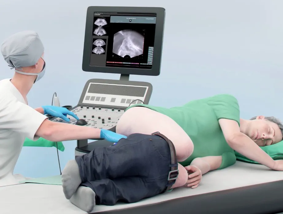
MRI fusion biopsy of the prostate is a targeted prostate biopsy technique that combines MRI imaging with ultrasound guidance to accurately sample suspicious prostate lesions. It is commonly used in patients with high PSA levels or abnormal prostate MRI findings to improve prostate cancer detection.
If you have been told that your PSA (Prostate Specific Antigen) level is high, your doctor may recommend a prostate biopsy to determine whether prostate cancer is present. Traditionally, prostate biopsies were performed using an ultrasound guided TRUS prostate biopsy, where multiple samples are taken randomly from the prostate gland.
Today, medical technology has evolved significantly. One of the most advanced methods available is the MRI Fusion Biopsy of the Prostate, which combines high-resolution MRI imaging with real-time ultrasound guidance to precisely target suspicious areas inside the prostate.
At our center, we specialize in advanced prostate cancer diagnostics and provide MRI fusion biopsy for patients with high PSA or suspicious MRI findings, helping achieve a more accurate diagnosis while minimizing unnecessary procedures.
Understanding PSA and Why Biopsy May Be Needed
PSA, or Prostate Specific Antigen, is a protein produced by prostate cells and measured through a blood test. PSA testing is widely used to screen for prostate diseases, including prostate cancer.
However, a high PSA does not automatically mean cancer. PSA levels can increase due to several conditions, including:
- Benign Prostatic
- Hyperplasia (BPH)
- Prostate inflammation or infection
- Recent ejaculation or catheterization
- Urinary tract infections
- Prostate cancer
When PSA levels are persistently elevated or rising over time, doctors often recommend further evaluation with MRI of the prostate and a prostate biopsy.
A biopsy remains the only definitive way to diagnose prostate cancer, as it involves examining small samples of prostate tissue under a microscope.
What is a Prostate Biopsy?
A prostate biopsy is a medical procedure in which small samples of tissue are taken from the prostate gland and sent to a pathology laboratory for examination.
The purpose of the biopsy is to determine:
- Whether prostate cancer is present
- The aggressiveness of the cancer (Gleason score)
- The amount and location of cancer within the prostate
Historically, most biopsies were performed using ultrasound guided TRUS prostate biopsy.
Ultrasound Guided TRUS Prostate Biopsy
The TRUS prostate biopsy (Transrectal Ultrasound-guided biopsy) has been the standard diagnostic method for many years.
During this procedure:
- An ultrasound probe is inserted into the rectum
- The prostate gland is visualized using ultrasound
- Multiple tissue samples (usually 10–12 cores) are taken from different regions of the prostate
Although TRUS biopsy is widely used and effective, it has some limitations.
Limitations of TRUS Prostate Biopsy
Because ultrasound images cannot always clearly identify cancerous areas, TRUS biopsies are often systematic or random. This means that the biopsy needle samples multiple areas of the prostate even if the suspicious lesion cannot be directly seen.
This approach can sometimes lead to:
- Missing clinically significant cancers
- Detecting low-risk cancers that may not require treatment
- Repeat biopsies if PSA continues to rise
To overcome these limitations, modern urology has adopted MRI fusion biopsy of the prostate
What is MRI Fusion Biopsy of the Prostate?
MRI Fusion Biopsy of the Prostate is an advanced diagnostic technique that combines:
- Multiparametric MRI imaging
- Real-time ultrasound guidance
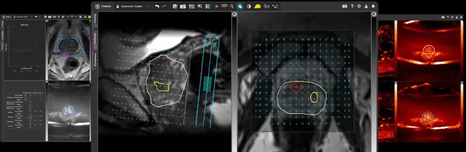
The MRI scan identifies suspicious areas inside the prostate, and specialized software fuses the MRI images with live ultrasound images during the biopsy procedure.
This allows the urologist to precisely target abnormal regions rather than sampling randomly.
As a result, MRI fusion biopsy improves the detection of clinically significant prostate cancer while reducing unnecessary sampling
Why MRI Fusion Biopsy is Recommended for Patients with High PSA
Patients with high PSA levels often undergo MRI before biopsy. If the MRI reveals suspicious lesions (usually reported using the PI-RADS scoring system), targeted biopsy becomes possible.
MRI fusion biopsy is particularly useful in the following situations:
- Persistently high PSA
- Suspicious lesions on prostate MRI
- Previous negative prostate biopsy with rising PSA
- Patients on active surveillance for prostate cancer
- Suspicion of anterior prostate tumors that may be missed by standard
TRUS biopsy
By targeting suspicious areas, MRI fusion biopsy improves diagnostic accuracy and reduces the risk of missing aggressive cancers.
How MRI Fusion Biopsy Works
The process of MRI fusion biopsy of the prostate involves several steps.
1. Multiparametric MRI Scan
Before the biopsy, the patient undergoes a multiparametric MRI (mpMRI) of the prostate. This imaging technique provides detailed information about prostate anatomy and helps identify suspicious lesions.
The MRI report usually assigns a PI-RADS score:
- PI-RADS 1 – Very low suspicion
- PI-RADS 2 – Low suspicion
- PI-RADS 3 – Intermediate suspicion
- PI-RADS 4 – High suspicion
- PI-RADS 5 – Very high suspicion
Lesions with PI-RADS 4 or 5 often require targeted biopsy.
2. MRI-Ultrasound Image Fusion
During the biopsy procedure, specialized software merges the previously obtained MRI images with real-time ultrasound images.
This process is called image fusion, allowing the urologist to see exactly where the suspicious lesion lies within the prostate.
3. Targeted Tissue Sampling
Using this fused imaging guidance, the biopsy needle can be directed precisely to the abnormal area.
Typically:
- 2–4 targeted cores are taken from the suspicious lesion
- Additional systematic cores may also be taken for comprehensive sampling
This approach combines the strengths of both targeted biopsy and systematic TRUS biopsy.
Advantages of MRI Fusion Biopsy of the Prostate
MRI fusion biopsy offers several advantages over traditional ultrasound guided TRUS prostate biopsy.
Higher Detection of Significant Cancer
MRI fusion biopsy improves the detection of clinically significant prostate cancer, which is the type that requires treatment.
Reduced Detection of Low-Risk Cancer
Because the biopsy targets suspicious areas, there is less chance of diagnosing insignificant cancers that may never cause harm.
Better Accuracy
By precisely targeting lesions identified on MRI, this technique improves diagnostic accuracy.
Useful After Previous Negative Biopsy
For patients with high PSA and previous negative TRUS biopsy, MRI fusion biopsy can detect cancers that were missed earlier.
Helps Guide Treatment Decisions
Accurate diagnosis allows doctors to determine whether a patient needs:
- Active surveillance
- Surgery
- Radiation therapy
- Other treatments
The Procedure: What Patients Can Expect
The MRI fusion biopsy procedure usually takes about 20–30 minutes.
Before the Procedure
Patients may be advised to:
- Stop certain medications such as blood thinners
- Take prescribed antibiotics
- Fast for a few hours if sedation is planned
During the Procedure
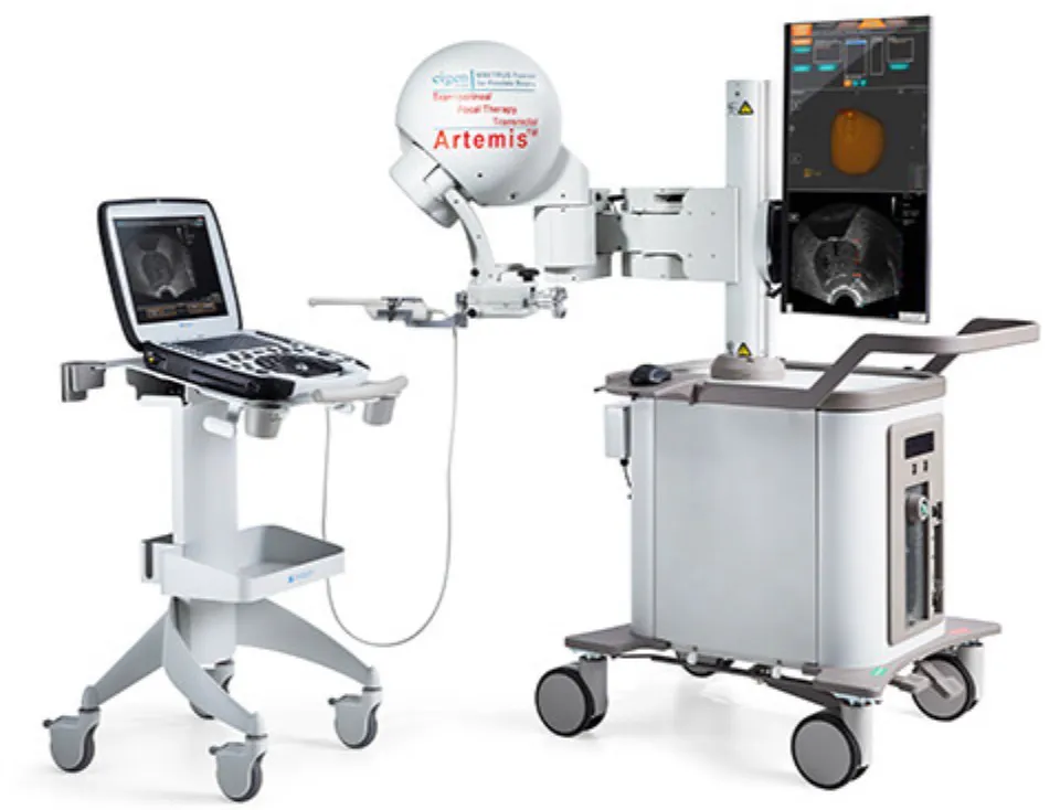
The procedure is typically performed under local anesthesia or mild sedation.
Steps include:
- Ultrasound probe placement
- MRI-ultrasound image fusion
- Targeted biopsy of suspicious lesions
- Systematic sampling if required
Most patients tolerate the procedure well.
After the Procedure
Patients can usually go home the same day.
Mild symptoms may occur, including:
- Blood in urine
- Blood in semen
- Mild rectal bleeding
These symptoms typically resolve within a few days.
Risks of Prostate Biopsy
Although prostate biopsy is generally safe, potential risks include:
- Infection
- Fever
- Urinary retention
- Temporary bleeding in urine or semen
Following proper antibiotic protocols significantly reduces infection risk.
Patients should contact their doctor if they develop:
- High fever
- Difficulty passing urine
- Severe bleeding
Who Should Consider MRI Fusion Biopsy?
MRI fusion biopsy may be recommended for patients with:
- Persistently high PSA levels
- Suspicious MRI findings
- Previous negative ultrasound guided TRUS prostate biopsy
- Rising PSA during active surveillance
- Suspicion of aggressive prostate cancer
Your urologist will evaluate your PSA trend, MRI findings, age, and overall health before
recommending biopsy.
Cost of MRI Fusion Biopsy in India
The cost of MRI fusion biopsy of the prostate in India varies depending on the hospital, imaging technology, and biopsy system used.
Although it is generally more expensive than traditional TRUS biopsy, the improved diagnostic accuracy may reduce the need for repeat procedures.
India is increasingly recognized as a global destination for advanced prostate cancer diagnosis and treatment due to:
- Experienced urologists
- Modern diagnostic technology
- Affordable healthcare costs
Frequently Asked Questions
Is MRI fusion biopsy better than TRUS biopsy?
MRI fusion biopsy is more accurate in detecting clinically significant prostate cancer compared to standard TRUS biopsy. However, many experts recommend combining targeted biopsy with systematic sampling.
Is prostate biopsy painful?
Most prostate biopsies are performed under local anesthesia, and patients usually experience only mild discomfort.
Can MRI miss prostate cancer?
Yes. Although MRI is highly useful, some cancers may not be visible on MRI. This is why biopsy remains the definitive diagnostic test.
How long does recovery take after biopsy?
Most patients return to normal activities within 24–48 hours.
When will biopsy results be available?
Biopsy results are usually available within 5–7 days, depending on the pathology laboratory.
Conclusion
For patients with high PSA or suspicious prostate MRI findings, accurate diagnosis is critical. Traditional ultrasound guided TRUS prostate biopsy has served as the standard method for many years, but modern advances such as MRI fusion biopsy of the prostate have significantly improved the precision of prostate cancer detection.
By combining advanced MRI imaging with real-time ultrasound guidance, MRI fusion biopsy allows targeted sampling of suspicious areas, improving diagnostic accuracy while reducing unnecessary procedures.
If you have been advised to undergo a prostate biopsy due to high PSA, consulting an experienced prostate cancer specialist can help determine whether MRI fusion biopsy is the right option for you.
Early and accurate diagnosis plays a crucial role in achieving the best possible outcomes for prostate cancer patients.